Expansive laboratory resources housed within the Department of Investigative Medicine at WMU Homer Stryker M.D. School of Medicine (WMed) are providing investigators opportunities to perform experiments that can potentially drive their research into unexplored territory.
Developed and built with collaboration and accessibility at the forefront, WMed’s Core Facilities offer state-of-the-art technologies to investigators, providing access to well-maintained shared instruments. The Core Facilities include a Flow Cytometry and Imaging Core, a Sequencing Core, and four Core Laboratories — an Equipment Laboratory, Tissue Culture Laboratory, Biosafety Level 2 Laboratory, and Radioisotope Laboratory.
“These facilities are incredibly valuable,” said Core Laboratory Manager Cody Jones, who maintains the labs, assisting with instrument training, setup, and use. “There are so many resources available, and in some instances, the equipment in our core labs serves as a valuable backup for equipment in the labs of our investigators, eliminating downtime in the case of equipment failure.”
Nichol Holodick, PhD, associate professor in the Department of Investigative Medicine, researcher in the Center for Immunobiology, and director of the Flow Cytometry and Imaging Core, said the core facilities have helped WMed be successful in obtaining extramural grant support for projects.
“If we did not have this infrastructure, it would be impossible to do things that we need to do to advance our research,” Dr. Holodick said. “The work that Cody Jones and our senior manager of the Flow Cytometry and Imaging Core, Michael Clemente, MS, SCYM (ASCP), do is so important. They keep all of the equipment in top shape and offer assistance wherever it’s needed.”
The following is a brief outline of each core facility:
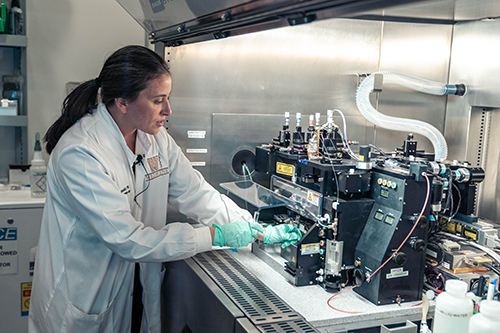
Flow Cytometry & Imaging Core
Flow cytometry is a lab technique used to measure the physical and chemical properties of cells by analyzing their light scattering and fluorescence emission characteristics.
At WMed, the Flow Cytometry and Imaging Core provides state-of-the-art equipment and expertise in flow cytometry, cell sorting, and imaging. Clemente, a flow cytometrist with extensive experience in immunology and hematology/oncology research, is readily available for project/experimental design consultation, data analysis, instrument setup, full-service cell sorting, and training.
“The flow core is an essential component of any immunology program, any hematology program, and has benefits for almost any cell study that people are doing right now,” Clemente said. “There's flow cytometry analysis, which involves essentially looking at cells or particles to determine their phenotype or their function. Then there's also flow cytometry sorting or what people refer to as fluorescence activated cell sorting. In that scenario, you're combining elements of physics and elements of biology with advanced computing power to sort cells or particles into plates or tubes at the single cell level.”
The recent addition of a cutting-edge spectral sorter and a cutting-edge spectral analyzer to the flow core has greatly expanded capabilities for investigators, allowing them to gain more information from fewer cells -- with the ability to apply/view up to 50 fluorescent tags simultaneously.
“It's really opened up whole new worlds in terms of the depth of knowledge that we can gain as to where a cell is going, where it might be coming from, what it's doing, what its role is, what type of receptors it has on its cell surface for chemotaxis or for activation or any of these different types of markers,” Clemente said. “The technology also allows you to not only identify the different properties of cells but then sort them into pure populations of the cells of interest. So if you're interested in, say, a B cell that has an activated phenotype, you can sort that cell very specifically using a set cluster of differentiation markers to identify what type of cell it is, and then also separate that out from the heterogeneous mixture of cells that you would find in tissue or in blood, or in cell cultures or any type of various scenarios you run into in science research.”
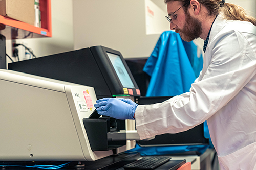
Sequencing Core
The Sequencing Core features a variety of DNA sequencing instruments, including a Sanger sequencer and an Illumina MySeq, allowing investigators to see exactly the order, or sequence, of nucleotides that make up particular DNA or RNA.
“We’re particularly interested in the DNA sequence itself, because — depending on the question being asked — it can yield different insights,” Dr. Holodick said.
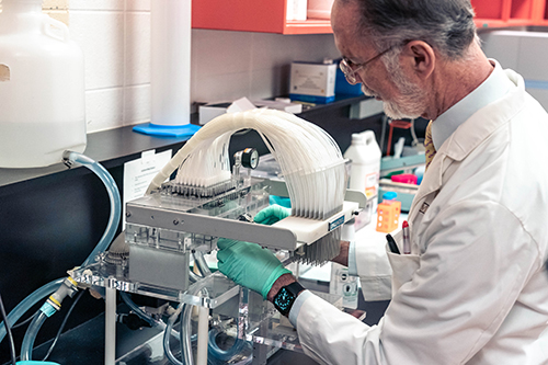
Equipment Laboratory
The Equipment Laboratory provides a wide variety of general resources, including plate readers, ultracentrifuges, PCR systems, and various analyzers.
“There are many tools in this lab to help investigators with their research,” Jones said. “Investigators are using the equipment to gather information to make discoveries and further their research.”
Tissue Culture Laboratory
The Tissue Culture Laboratory provides ample resources for researchers who need to work with cells in a sterile, controlled setting. The facility allows the culture of human and other eukaryotic cells.
“The Tissue Culture lab provides a controlled environment to grow cells outside of the body,” Dr. Holodick said. “We’re trying to replicate physiological conditions in the body to keep the cells alive, which enables us to study the cells’ behavior.”
Biosafety Level 2 Laboratory
The Biosafety Level 2 Laboratory offers a shared dedicated space to perform investigative techniques requiring an elevated level of containment, such as bacterial work.
Residing in a separate room under negative pressure, the Biosafety Lab was built to BSL2 specifications. Researchers are required to undergo specific training and certification prior to obtaining access to the lab, and required personal protective equipment (PPE) is provided.
“The lab includes freezers, biosafety cabinets, as well as incubators for growing bacteria, viruses, and cells,” Jones said. “Researchers work within the BL2 core because some research involves biological materials that pose a moderate risk to people. Therefore, the work has to be done in a secure and controlled space to avoid putting anyone at risk.”
Radioisotope Laboratory
The Radioisotope Laboratory resides in a separate dedicated room with all necessary equipment for work with radioactive material commonly used in biological research, such as Tritium, a radioactive isotope of Hydrogen.
Researchers are required to undergo training and certification prior to obtaining access to the lab, and required personal protective equipment (PPE) is provided.
"I use tritiated thymidine, which is a radioactive form of the DNA building block thymidine, to measure cell division,” Dr. Holodick said. “For example, I take cells from a mouse or a human and culture them with bacterial components. The B cells, a type of immune cell that makes antibodies, recognize these components and divide, producing more cells. Often, I want to see whether factors like hormones affect their ability to divide in response to these bacterial components. By adding tritiated thymidine — a radioactive label — the only cells that incorporate it into their DNA are those actively dividing. We can then measure the incorporated radioactivity to quantify cell division. A stronger signal means more cells were dividing, while a weaker signal means fewer were dividing."
WMed’s core facilities have been developed, outfitted and enlarged over the past nine years through the efforts of members of the Center for Immunobiology and the Department of Investigative Medicine, and through support provided by the medical school. Prior to 2016, none of this infrastructure existed at the medical school.
Today, the core facilities are available for use by any WMed investigator. These facilities — particularly the Flow Cytometry and Imaging Core — have also become regional resources for biomedical investigation in Southwest Michigan, drawing in investigators outside of WMed.
“We hope that in this way we can advance research here that changes the way medicine is practiced to more effectively counteract disease and alleviate suffering,” said Thomas L. Rothstein, MD, PhD, chair of the Department of Investigative Medicine and director of the Center for Immunobiology at WMed.
For more information about WMed’s Core Facilities, visit wmed.edu/investigativemedicine/corefacilities.