A hidden gem tucked within the Department of Pathology at Western Michigan University Homer Stryker M.D. School of Medicine (WMed) is helping provide timely answers to public health partners, researchers and families alike.
The Research Histology Laboratory, located on the fourth floor of the W.E. Upjohn M.D. Campus in downtown Kalamazoo, provides an array of routine and specialized histological services and immunohistochemical staining for autopsy, veterinary, and research specimens.
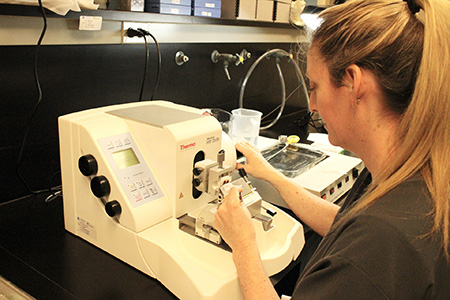
“We handle all of the tissue processing. Once tissue samples are obtained from autopsies, neuropathology examinations, external client projects, or the faculty labs that adjoin us, those all come into our lab and are processed,” said Kristi Bailey, HTL, lead histotechnologist at WMed. “Our variety of stains highlight the tissue components on a microscopic level, so that the samples can be viewed under the microscope. Some of the specialty stains we use will highlight particular tissue components, allowing pathologists to see what's going on within those tissues.”
Services offered by the Research Histology Lab include:
- Tissue processing
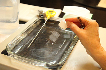
- Slide preparation from paraffin-embedded and frozen tissues
- Bone decalcification and sectioning
- Unstained slide preparation, serial and step sectioning
- Routine and specialized histological staining
- Immunohistochemical staining
- Digital imaging of slides
Launched in 2016, the Research Histology Lab has the capability to process a wide variety of samples, from small samples of tissue to much larger, fully mounted brain sections. The lab processes roughly 11,000 samples per year, providing key support to WMed’s Office of the Medical Examiner and Forensic Services – which serves as the medical examiner for 13 counties in Michigan – and the Division of Neuropathology, among others.
“We cannot do what we do that without histology, it will not work,” said Amanda Fisher-Hubbard, MD, vice chair of the Department of Pathology and chief of the Division of Neuropathology. “Our histology lab is one of the reasons I’m here at WMed, because we have such great resources.”
The Division of Neuropathology regularly receives autopsy brains, eyes, spinal cords, and cervical spines recovered from decedents with histories of seizures, cerebral palsy, dementias, and traumatic head injuries, including abusive head trauma. The Research Histology Lab offers specialized histologic stains, as well as immunohistochemical stains, to aid in interpretation and diagnosis. Additionally, the lab serves as a collection site for the Lieber Institute for Brain Development, helping support their research endeavors.
“What we've created here is done so well and managed so well, it’s a highlight of what we do and it’s such an impressive piece of what we do,” Abigail Grande, senior manager of research and autopsy operations at WMed, said of the Research Histology Lab. “It's such a vital part of a medical examiner’s function to have this at our fingertips, and to be an academic medical examiner's office where we are embedded in the medical school ... Most medical examiner’s offices cannot do quite the level of academic research that we can.”